INDICATIONS:
Medical Necessity: Evaluate for gallstones and dilated common bile duct and sonographic signs or cholecystitis, such as “positive sonographic Murphy’s sign,” thickened gall bladder wall, and pericholecystic fluid
Structures studied: Gallbladder (GB), common bile duct
REQUIRED IMAGES:
Short Axis, Long Axis, Common Bile Duct
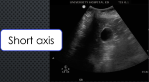
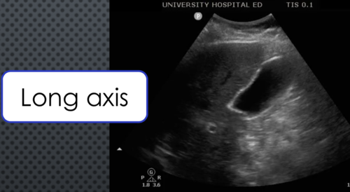
1.) Obtain a video clip of the GB in it’s entirety, in both the longitudinal and transverse planes.
2.) Obtain a static image of the GB in the transverse plane with a measurement of the anterior GB wall.
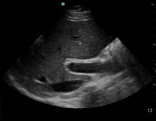
TRANSVERSE VIEW OF GALLBLADDER
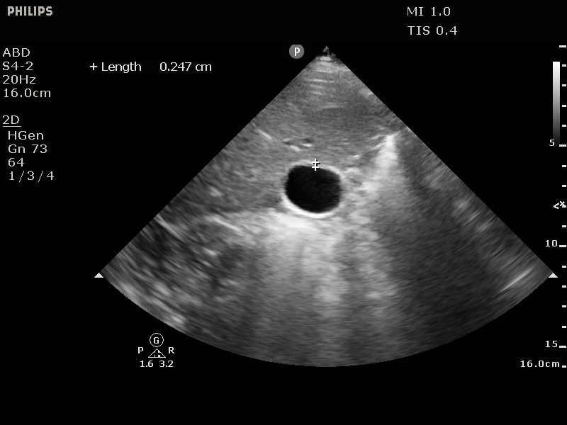
ANTERIOR WALL MEASUREMENT IN THE TRANSVERSE PLANE
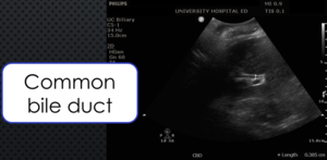
3.) Obtain a static image of the Common bile duct (CBD).
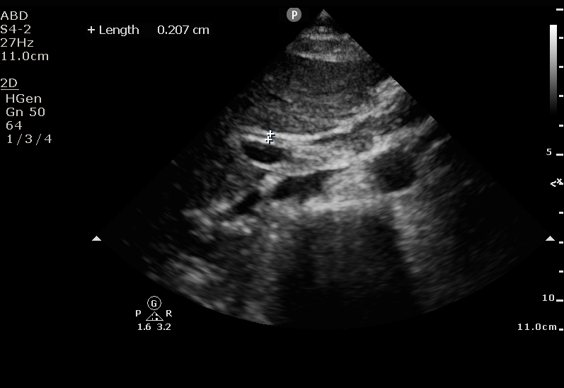
COMMON BILE DUCT MEASUREMENT
CRITICAL MEASUREMENTS
COMMON BILE DUCT
<5mm-normal in patients up to age 50 (Add 1 mm for every decade of life thereafter)
5-7mm equivocal
>7mm-dilated
GALLBLADDER WALL
<3mm-normal
3-4mm-equivocal
>4mm-dilated
Note: The left lateral decubitis, in addition to the supine position, is generally superior for the GB and CBD visualization.


