The Abdominal XRay: A relic or a reliable tool?
/The KUB is easy to obtain and commonly used in the Emergency Department despite the advent and ease of higher specificity radiological studies. As these studies are still readily used and in some settings may be the only imaging available, our goals for this post will be to discuss indications for abdominal radiography in the emergency department as well as give examples of normal anatomy, an approach to interpretation, and underlying pathology that can be discovered, including associated sensitivities and specificities. We will not discuss utilization of contrast. Furthermore, we will briefly touch on different abdominal views that can be obtained and their utility but will focus primarily on the KUB since it is the most utilized image obtained in the emergency department.
Overview
ACR Indications for KUB
- Evaluation and follow-up of abdominal distention, bowel obstruction, or nonobstructive ileus
- Constipation, especially assessment of fecal load in children
- Evaluation for necrotizing enterocolitis, particularly in the premature newborn
- Evaluation of congenital gastrointestinal abnormalities
- Follow-up of the postoperative patient, including detection of inadvertent retained surgical foreign bodies
- Evaluation and follow-up of urinary tract calculi, including assessment of lithotripsy patients
- Evaluation of ingested or other introduced foreign bodies
- A scout radiograph prior to a planned imaging examination, ie, fluoroscopy
- Evaluation of the placement of medical devices
- Evaluation for pneumoperitoneum
- Evaluation of possible toxic megacolon
- Evaluation of unstable patients after blunt trauma to the abdomen
- Evaluation of a palpable mass in an infant or child
- Localization of pancreatic duct stone pre-lithotripsy and endoscopic stone removal
- Evaluation for suspected retained video endoscopy capsule and determination of location of patency capsule
- Evaluation of colon transit time using the simplified radiodense marker colon transit test
There has been constant debate surrounding the indications and utility of abdominal plain imaging. The American College of Radiology have listed the 16 indications for abdominal radiography (table at right). Realistically the abdominal radiograph (AXR) should be ordered when there will likely be a finding for a suspected disease process and the imaging will change management. Rothrock, Green, and Hummel found the presence of prior abdominal surgery, foreign body ingestion, abnormal bowel sounds, abdominal distention, or peritoneal signs was 93% sensitive in detecting diagnostic radiographs in children presenting with major diseases.
Plain abdominal radiography is frequently over-ordered in the emergency setting and are often the only imaging study obtained (Greene Indications for plain AXR). This increases healthcare cost and unnecessary radiation to the patient. The radiation exposure for an AXR 1.2-1.5 mSv exposure per abdominal film which can equate to 75 chest x-rays. This is not negligible when increased cancer risk may occur between 10 and 100 mSv. The most common view of the abdominal radiograph is the anteroposterior projection in the supine position. The practitioner should start with a KUB and possibly an upright chest x-ray and obtain additional radiographs as indicated.
Technique
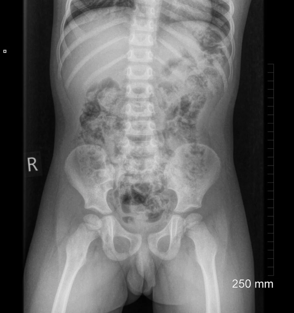
Supine
Patient supine on table with the whole diaphragm visualized superiorly and the inferior pubic rami visualized inferiorly. The lateral abdominal wall musculature and soft tissue should be visualized.
Case courtesy of Dr Aneta Kecler-Pietrzyk, Radiopaedia.org, rID: 53076
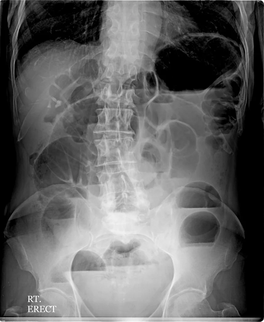
Upright
This is performed with the patient standing upright. Here visualization of air-fluid levels are more easily identified. This view is indicated when an obstruction or large gas-producing abscess is suspected. The upright x-ray is not ideal for detecting a small pneumoperitoneum.
Case courtesy of Dr Ahmed Abdrabou, Radiopaedia.org, rID: 35721

Lateral
Decubitus
This view is obtained in the left lateral decubitus positon (the right lateral decubitus is rarely helpful). This is more sensitive for identification of small pneumoperitoneum or obstruction, especially when the patient is too ill to stand. Lastly it may better reveal any intussusception given the better image of the right lower quadrant.
Case courtesy of Dr Maulik S Patel, Radiopaedia.org, rID: 13853
Other Views: Only indicated if pathology found on initial imaging and when other imaging modalities are not indicated or not available.
- Erect Chest Film- Evaluates for free air or underlying chest pathology. Please refer to Dr. Alexa Sabedra’s post on Interpreting Chest X-rays.
- Prone view for suspected anorectal malformations, intussusception, or intraabdominal abscess.
- Cross-table supine to evaluate for foreign object in penetrating trauma. May have increased sensitivity in neonates for detecting free air (ACR)
- Collimated low kilovoltage to evaluate for a specific area of interest for free air, stones, or abscess.
- Oblique view may reveal calcifications that were obscured by the spine.
Approach
In order to accurately interpret the AXR the practitioner must follow a repeatable step-by-step approach every time. This ensures that small findings are not missed especially in the presence of huge abnormalities. We will discuss the easy to remember: “Gasses, masses, bones, stones, leads, and lines”
- Gasses- Evaluate the bilateral lungs bases, the bowel gas pattern, evaluate for any sings of air outside of the bowel including pathologic air in the solid organs or subcutaneous tissue.
- Masses- Review the solid intra-abdominal organs looking for any abnormalities in size, outlines, blurring, or mass effect.
- Bones- Evaluate the visualized skeleton for an evidence of fracture, malalignment, or abnormal lesions.
- Stones- Look for any calcifications or ingested radiopaque objects
- Leads and Lines- Evaluate for any tubes, lines, or drains for appropriate placement.
Pathology
Malrotation with volvulus
Conventional radiography is not sensitive nor specific for malrotation. Findings on AXR may include air-fluid levels consistent with obstruction, dilated loops overlying the liver, or potentially a double bubble sign. Furthermore, AXR may show right-sided jejunal marking with absence of stool filled colon in the right lower quadrant. Interestingly, normal gas pattern is the most common finding in malrotation. Ultimately plain radiography should not be used to exclude malrotation and volvulus.
Study of choice - upper GI series
Necrotizing Enterocolitis
AXR is the imaging modality of choice. When NEC is suspected obtain AP and lateral decubitus abdominal radiographs. Repeat imaging can be performed every 6 hours to evaluate for rapid evaluation. Findings are on a spectrum that change with disease severity. The table below is a standard 10-point scale that increases with disease severity and for every 1-point increase patient is more likely to have severe disease measured by need for surgical intervention. Both Bell’s criteria and modified Bell’s criteria follow a similar trend of ileus -> intestinal pneumatosis -> Portal venous gas -> ascites -> pneumoperitoneum. Pneumatosis intestinalis is highly specific for NEC and present in 75% of cases. Portal venous gas is present in 10-30% of cases.
Pneumoperitoneum in NEC is associated with several signs. These include Rigler’s sign, visualization of intraabdominal ligamentous structures, and football sign. Hover over the images below for the specific sign and severity of the NEC.

Intussusception
For ileocolic intussusception, sensitivity of AXR is 74 to 90% and approaches 100% sensitivity if there is air present in the ascending colon in three separate views. However, up to 24% of children with intussusception may have normal radiographs. The practitioner should focus on the cecum and the entire large bowel. However, children occasionally have redundant sigmoid colon that may extend to the right lower quadrant and be mistaken for the cecum. Prolonged intussusception may lead to signs of bowel obstruction and perforation. Ultrasound is the imaging modality of choice but if radiography is attempted, two views should be utilized. Signs consistent with intussusception include Crescent Sign, Meniscus Sign, and Target sign (pictured below). Of note, this is the most common cause of SBO in children.
Appendicitis
AXR is rarely useful and may reveal a appendicolith which is highly suggestive of appendicitis but only visualized in 5-10% of cases. Other findings include focal right lower quadrant ileus, loss of psoas shadow, or right lower quadrant soft tissue mass. Lastly the inflammation may irritate the ipsilateral psoas muscle leading to muscle spasm and scoliosis convex to the left side. None of these are definitive for appendicitis.
Bowel Obstruction
AXR is 30-70% sensitive and 50% specific for obstruction and can reveal dilated loops of bowel with air-fluid levels. Emergency Department providers had a sensitivity of 47% and a specificity of 72% when detecting SBO on supine AXR. When a upright or decubitus view those numbers rose to 69% and 73% respectively.
Constipation
AXR is often ordered for constipation and only have a sensitivity of 60-80%. Misdiagnosis in children with constipation is more frequent in patients where an AXR was obtained and patients with abdominal pain and tenderness. The most common missed diagnosis included perforated appendicitis, intussusception, and bowel obstruction. Review of literature revealed no correlation of clinical constipation and fecal load appreciated on AXR. AXR has little or no role in confirming or ruling out a diagnosis of constipation. Therefore despite a large stool burden on AXR clinicians should not discount serious/alternate causes of abdominal pain.
Renal Stones
AXR can detect up to 90% of renal calculi although stool may make this more diicult to view. Radiography has a sensitivity of 62 % and specificity of 67 % of detecting stones. They have no advantage over CT scan and should not be ordered instead of CT. Calcium stones are radiodense. Struvite somewhat radiodense and may show up. Uric acid and cysteine are radiolucent.
Cholelithiasis
AXR can detect 15% of gallstones in adults. However, given the pathophysiology of gallstone formation in children (hemolytic disease, TPN, and cystic fibrosis) they may be seen 50% of children.
Tumors
Any masses appreciated on physical examination should begin evaluation with ultrasound. However occasionally AXR is ordered and a mass may be noticed. Calcification is present in at least 30% of neuroblastomas and this may been seen on AXR.
Inguinal hernia
May be missed on AXR if the image does not include the scrotum on imaging.
Inflammatory Bowel Disease
Children account for one-quarter of new diagnosis of IBD. AXR can reveal classic thumbprinting and may be used to evaluate for toxic megacolon
Non-accidental Trauma
AXR may reveal evidence of abuse. This is why it is important to make sure you evaluate everything in the image and not just what is in the abdomen.
Post by Trevor Skrobut, MD
Editing by Ryan LaFollette, MD
References
- Almaramhy, Hamdi Hameed Acute appendicitis in young children less than 5 years: review article, Italian Journal of Pediatrics, 10.1186/s13052-017-0335-2, 43, 1, (2017).
- Marx, John A., and Peter Rosen. Rosen's Emergency Medicine: Concepts and Clinical Practice. 8th ed. Philadelphia, PA: Elsevier/Saunders, 2014
- Pickhardt, Perry J. and Bhalla, Sanjeev Intestinal Malrotation in Adolescents and Adults: Spectrum of Clinical and Imaging Features American Journal of Roentgenology. 2002;179: 1429-1435. 10.2214/ajr.179.6.1791429
- Applegate KE (2009) Evidence-based diagnosis of malrotation and volvulus. Pediatr Radiol 39(Suppl 2):S161–S163
- Strouse PJ. Malrotation. Semin Roentgenol 2008; 43:7–14
- Tintinalli, Judith E.,, et al. Tintinalli's Emergency Medicine: A Comprehensive Study Guide. Eighth edition. New York: McGraw-Hill Education, 2016
- Coursey CA, Hollingsworth CL, Wriston C et al (2009) Radiographic predictors of disease severity in neonates and infants with necrotizing enterocolitis. AJR Am J Roentgenol 193:1408–1413
- Smith J, Fox SM. Pediatric abdominal pain. An emergency medicine perspective. Emerg Med Clin North Am. 2016;34:341–61
- Elsayes, Khaled and Oldham, Sandra. Introduction to Diagnostic Radiology. New York: McGraw-Hill Education, 2014.
- Sleisenger, Marvin H, Mark Feldman, Lawrence S. Friedman, and Lawrence J. Brandt. Sleisenger and Fordtran's Gastrointestinal and Liver Disease: Pathophysiology, Diagnosis, Management. Philadelphia: Saunders/Elsevier, 2010.
- James B, Kelly B. The abdominal radiograph. Ulster Med J. 2013;82:179–187
- CURRENT Diagnosis & Treatment: Emergency Medicine, 8e Eds. C. Keith Stone, and Roger L. Humphries. New York, NY: McGraw-Hill, ,
- Freedman SB, Thull-Freedman J, Manson D, et al Pediatric abdominal radiograph use, constipation, and significant misdiagnoses. J Pediatr 2014; 164:83–88. e82.
- Saps, M., Rosen, J.M., Ecanow, J. X-ray detection of ingested non-metallic foreign bodies. World J Clin Pediatr. 2014;3:14–18.
- Florez MV, Evans JM, Daly TR (1998) The radiodensity of medications seen on X-ray films. Mayo Clin Proc 73:516–519
- Sarah J. Menashe, Ramesh S. Iyer, Marguerite T. Parisi, Randolph K. Otto, Edward Weinberger, and A. Luana StanescuPediatric Abdominal Radiographs: Common and Less Common Errors American Journal of Roentgenology 2017 209:2, 417-429
- Beinvogl, Beate et al. Are We Using Abdominal Radiographs Appropriately in the Management of Pediatric Constipation? The Journal of Pediatrics , Volume 191 , 179 - 183
- Daneman A, Navarro OM. Common pitfalls in paediatric abdominal imaging. Pediatr Radiol 2009; 39(suppl 3):369–371
- Musson RE, Bickle I, Vijay RKP Gas patterns on plain abdominal radiographs: a pictorial review Postgraduate Medical Journal 2011;87:274-287
- Greene CS . Indications for plain abdominal radiography in the emergency department. Ann Emerg Med 1986;15:257–60
- Rothrock, Steven G et al. Plain abdominal radiography in the detection of major disease in children: A prospective analysis Annals of Emergency Medicine , Volume 21 , Issue 12 , 1423 – 1429
- Fleisher, Gary et al. Interpretation of pediatric x-ray films by emergency department pediatricians Annals of Emergency Medicine , Volume 12 , Issue 3 , 153 – 158
- Miele V., Piccolo C.L., Saracco V., Napoletano M., Trinci M., Brunese L. (2016) Imaging Non-traumatic Abdominal Emergencies in Pediatric Patients. Springer, Cham
- Hughes, U.M., Thomas, K.E., Shuckett, B. et al, The abdominal radiographic series in children with suspected bowel obstruction—Should the second view be abandoned?. Pediatr Radiol. 2002;32:556–560.
- https://radiopaedia.org
- https://www.radiologymasterclass.co.uk/https://www.acr.org/-/media/ACR/Files/Practice-Parameters/radabd.pdf?la=en
- Berrocal T, Lamas M, Gutiérrez J, Torres I, Prieto C, del Hoyo ML. Congenital anomalies of the small intestine, colon, and rectum. RadioGraphics 1999; 19:1219–1236


