Grand Rounds Recap 9.21.2016
/CNS Devices & Complications with Dr. Owens
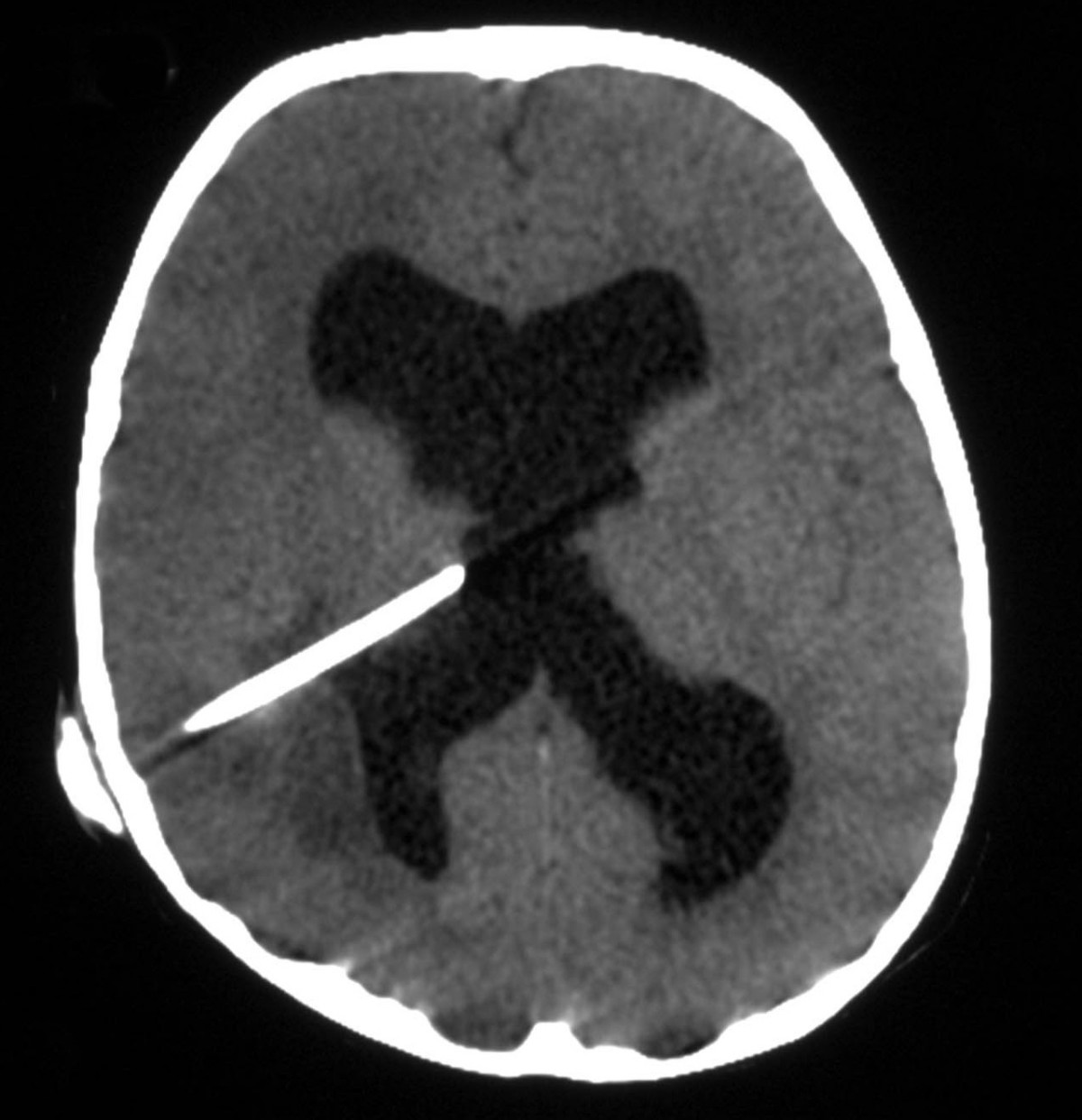
Ventriculoperitoneal Shunts: placed for hydrocephalus with one end in a ventricle, reservoir that sits under the skin on the skull and an end that drains distally into a receiving area, usually intra-abdominally
Symptoms of shunt complications
- AMS
- Changes in balance/coordination
- Seizure
- Incontinence
- Vision abnormalities
Shunt obstructions:
- Proximal obstruction: blockage of the end in the ventricle by debris, infection, or blood
- Often have a dry tap of the reservoir
- Distal obstruction: something at outflow tract is blocking outflow
- Often have a congested appearing shunt reservoir
Shunt Infections:
- Staph epidermidis in infections early after placement
- Staph aureus in delayed / late infections
Slit Ventricle Syndrome: Classic triad of symptoms of shunt obstruction + slit like ventricles on CT + slow filling of shunt reservoir
Baclofen Pumps: placed for spasticity
Oral Baclofen: need high doses with increased side effects and less of a reduction in muscle contractions, poor BBB penetrance
Intrathecal Baclofen Pump: much lower doses, less side effects, and increased efficacy
Baclofen withdrawal syndrome: Consider empty pump reservoir catheter leak/displacement, pump malfunction, refill with wrong drug concentration
- Symptoms: Hallucintations, psychosis, hyperthermia, rhabdo, cyclones, seizures, CID, organ failure, coma death
- Management: look around for pump site, benzos, benzos, benzos, oral baclofen, dantrolene, Call NSGY, imaging
Baclofen Toxicity: Consider pump malfunction, renal dysfunction (85% renally cleared)
- Symptoms: hypotonicity, hypothermia, bradycardia, hypo/hypertension, respiratory suppression, seizures, non-convulsive status
- Management: Look for a pump, supportive care: pressors, IVF dialysis
Spinal Cord Stimulators: last resort treatment for pain, first will have a trial with percutaneous electrodes and if the patient gets above a 50% pain improvement—> permanent implant.
- Complications: Overall complication rate 19.5%
- Hardware Failure w/o neurologic injury (38%), Infection (3.4-6%)—> remove electrodes
Varicella with Dr. Sabedra
Case: 1 year old with rash, fever, and RUE swelling for one week, diagnosed with primary varicella infection at OSH earlier in the week. Fevers >100 and RUE swelling that started the night of presentation. No injury to the arm, doesn’t want to move the arm. VS T 100.2, HR 160, RR 36 BP 118/89. Appears unwell but not toxic, no active ROM R arm, holds it flexed at the elbow, non-pitting swelling from the wrist to mid humerus, will allow for passive ROM. x-ray normal. WBC 34, ESR 62, CRP 19. The patient was admitted to medicine with vanc/ceftriaxone, hand called cellulitis and then got MRI for osteo. MRI positive for osteomyelitis, fluid collection in the forearm, joint fluid infected, group A strep grew from blood cultures. Went to OR found to have necrotizing fasciitis.
Chicken Pox / Varicella:
Transmission: 1-2 days prior to rash is when you start being contagious and continue to be contagious until lesions are crusted over. Infected secretions, fluid from rash, and can be airborne.
Vaccine for Chicken Pox: 1995, one dose schedule -->, 85% reductions in US cases and 88% reduction in varicella related hospitalizations. 2 doses in 2005--> still studying outcomes but > 95% reduction from initial population
Breakthrough varicella: infection after exposure in those who been vaccinated

Primary vaccine failure: failure to mount a protective immune response - 25% after first vaccine
Secondary vaccine failure: loss of immunity after initial vaccine response
1 year after - 97% effective
2-8 years after vaccine - 84% effective
No long-term data on duration of protection of the 2 dose regiment
Complications from Varicella infection: 90% admitted to hospital varicella described as healthy/immuocompetent
Neurological Complications:
Cerebellar ataxia (1/4000) direct infections of the cerebellum vs parainfectious immunological mediated demyelinating process, CSF most often normal, self-limited, no substantial evidence that antiviral therapy alters course
Varicella Encephalitis (1-2/10,000): CSF with increased opening pressures, mild-to-moderate lymphocytic pleocytosis (usually <100cells/uL), Mortality 5-10% with few poor outcomes otherwise
Pulmonary Complications:
Pneumonitis/pneumonia: increased frequency in immunocompromised adolescents and adults, diffuse interstitial nodular infiltrate, anecdotal reports suggest antiviral benefit without great data
Bacterial Super-infection:
Most common complication, break down in skin barrier gives access to bacteria; osteomyelitis, necrotizing fasciitis. 73% of hospitalized children had superficial infections , 27% deep infections
NSAIDs, Varicella & Nec Fasc: Use of NSAIDs in children with varicella might increase risk of soft tissue and invasive GAS infections, variable reports in literature biases by retrospective design
Ultrasound Guided Regional Anesthesia (USGRA): General Principles with Dr. Carleton
USGRA Challenges
- Room, patient and US-machine set up
- Small indistinct targets
- Asnisotropy
- Tissue mimics
- Complex local anatomy
- Maintaining in-plane needle visualization
- Injection visualization
Optimizing the Image

- Short axis views of the nerve provide best view
- Long axis views of needle provide best view
- Position the patient for comfortable access
- Position yourself for comfortable access
- Hold the probe in your non-dominant hand
- Hold the syringe/needle in your dominant hand
Pre-procedural Image Assessment
- Identify the target and surrounding structures to avoid
- Position the target on the screen to also allow for visualization of the needle path
- Scan the entire planned needle path
- Use the color doppler to identify vessels in the planned needle path or near the target
- Optimize image in terms of gain, TCG, focus, THI
Optimizing the image
- Imaging mode- compound imaging +/- tissue harmonic imaging (THI)
- Compound imaging: Multiple beams sent out by each crystal, improving lateral resolution (defining edges)
- THI: turn on for vascular imaging
- Depth- center target in image
- Frequency- probe selection and R(resolution) P (penetration) G (general) setting
- Focus- set beam focus at depth of target tissue
- Gain- overall + TGC at depth of target tissue
- Doppler - interrogate planned needle path
Needle Path
- Shallow allows for visualization of the needle throughout
- Practice walking the needle to the target
- Needle enhancement (MBE)
- Needle localization with hydrolocalization or tissue movement
Giving Feedback Small Groups with Dr. Paulsen

Learning is a function of feedback. Satisfaction is a function of praise.
Disconfirming: Below the level of what you perceive (only negative)
Confirming: at or above the level of what you perceive (only positive)
Discordant: does not confirm what you perceive
Concordant: confirms what you perceive
Tips for Feedback:
- Label Feedback
- Self-assessment
- Beware of overload (Pick one thing: Knowledge, translation, communication, procedural skill, attitude)
- Be specific and strategic
- Use a Feedback Tool
15% of medical students are difficult learners
- What is the problem?
- Whose problem is it?
- Should it be addressed? If so, then how?
Difficult Learner #1: Medical Knowledge Deficits
- The problem is there are medical knowledge deficits and also some skills of reporting
- Its a learner and system problem both, we may have failed the learner in teaching what was needed
- Yes. Give specifics about what to read, not just “read more” but give a certain paper. Support them looking for their own answers
Difficult Learner #2: Knowledge Translation Deficits
- Skill
- Learner>System>Teacher
- Yes. Use socratic method to probe into the thought process more concretely
Difficulty Learner #3: Attitude
- Attitude
- Learner/Teacher
- Yes. Give specifics about why the attitude affected the shift and give examples of why the professionalism is an issue
ED Super-utilizers with Dr. Betham
In patients with >20 visits v 6-20 visits, those with >20 visits...
- Are less sick when they show up
- Have less “appropriate use of the ED” (left ama more often)
- Have a lower admission rate
- Cost per visit was lower (793 v 2294)
Our Superusers: 20 visits or more + admission rate of 20% or had at least 12 admissions
- 25 patients in the group
- 60% female
- Admission rate 19-70%
- 717 visits/318 admissions
- 100% of them are insured
- 1/3 of the time they are here during office hours of their PCP
- Average acuity level 3
- 3-6 hours in the ED before disposition
- Spend a cumulative time of 231 days in the ED
- 65% of them go to med/surg beds
- 2/3rds of them stay longer than 2 midnights
- Total inpatient days 883
- 40% of the patients have no scheduled follow-up
- 52% of the follow-up appointments that are scheduled are attended
Case Management is the most effective, cost-neutral and does reduce ED utilization
Tactical Vent Management: The impact vent with Dr. Powell
Stealth mode- Menu and On button together
Starting the vent: plug green stuff into green stuff, clear stuff into clear stuff
There are baseline settings, turn it on and hook it up and you can do baseline things if you remember nothing else.
Volume Control
- Tidal volume amount is selected
- Volume is the constant set on the ventilator
Pressure Control
- Set pressures
- P delta drives tidal volume delivered= PIP-PEEP
- Decreasing P delta decreases TV
Pplat
- Chest wall compliance
- Lung compliance
- Pplat obtained currently with inspiratory pause
- Alveolar pressure
PIP
- Includes Pplat
- Also includes airway resistance
- Airway pressure
- Flow
- Resistance
- Alveolar pressure
If the Pplat is >30, consider:
- Mucus plug
- Kinked tubing
- Bronchospasm (check plateau pressure to make sure they are significantly different, can help with diagnosis)
Most patients will be on volume control while pressure may be more comfortable
Volume control to pressure control
- Calculate desired volume
- Adjust Pdelta to get volume
- Set PEEP and PIP to obtain Pdelta
ARDS
- ARDSNet study
- 8cc/kg to 6cc/kg down to as little as 4 cc/kg
- start with 6 cc/kg
Need to calculate if you will have enough oxygen until a new tank shows up?
- Cylinder Volume (L)= Tank Pressure (PSI) x Tank Factor
- D cylinder constant 0.16
- Calculate MV
- Calculate total L oxygen needed
- Calculate cylinder volume
- Divide L requirement on the vent by the cylinder volume


