Annals of B-Pod: Quick Hit Case
/Open Globe and a Discussion about Traumatic Hyphema
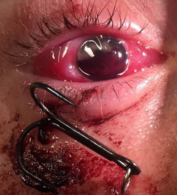
The patient with a fishhook penetrating his eye. Note the presence of a hyphema. Consent was given by the patient for the use of this image.
The patient is a male in his 40s who presents after sustaining an injury to his right eye with a fishing hook. He states that a three-barbed hook pierced his eye while fishing with his friend. On gross inspection, the hook was noted to have pierced the inferior eyelid causing an obvious right open globe and there was a large hyphema. While he was initially able to count fingers at four feet in his superior visual field, his visual acuity quickly deteriorated to light perception only. Extraocular movements were intact and caused movement of the hook. Ophthalmology was consulted and a CT was obtained. The patient was then taken to the OR for anterior chamber washout, open globe repair, and removal of the fish hook. He was discharged following the surgery with next day follow-up with ophthalmology.
Management and Evaluation of Traumatic Hyphemas and Open Globe Injuries
A hyphema, defines as a collection of blood in the anterior chamber, is most commonly caused by trauma to the eye. [1] Blunt trauma causes a hyphema by stretching or shearing anterior uveal structures. Penetrating trauma causes hyphema due to the direct vascular injury. Hyphema can also form spontaneously in conditions such as leukemia/lymphoma, ocular neoplasm, coagulopathies, and sickle cell disease. [2]
When evaluating a patient with a hyphema, a slit lamp exam should be performed to rule out a corneal abrasion, evaluate for traumatic iritis, and visualize the extent of bleeding and clot formation. Siedel’s test is useful to evaluate for open globe injury. To perform the test the clinician anesthetizes the globe with tetracaine and applies fluoroscein just as one would to evaluate for a corneal abrasion. The eye is then examined with a Woods lamp or the blue cobalt slit lamp filter. If perforation or leakage is present, the concentrated dye will be diluted by the aqueous humor from the anterior chamber.

CT orbit showing Entrance of the hook into the sclera, decreased size of the right globe, and a small hemorrhage anterior to the lens.
Only once an open globe injury is excluded should a clinician obtain intra-ocular pressures. Intra-ocular pressures are necessary because patient’s with hyphema are at high risk for secondary glaucoma due to blockage of aqueous humor drainage by clotted blood. [1] Patients with sickle cell disease are particularly susceptible to this pathology. [3] Management of hyphema focuses on preventing further trauma, preventing rebleeding, and treating complications. [3] An eye shield is typically applied to prevent further trauma. [3] The head of the bed should be raised to at least 30 degrees whenever the patient is supine to prevent secondary glaucoma caused by blood settling posteriorly. [3] A cycloplegic such as atropine can be used to immobilize the pupil to prevent further injury to vessels of the anterior chamber. [3] Topical steroids can be considered to prevent secondary uveitis associated with traumatic hyphema.3 Given the limited evidence to support these treatments, decisions to use these medications should be made on a case-by-case basis and in consultation with ophthalmology. [3] Antifibrinolytics such as oral aminocaproic acid have been shown to reduce the rate of rebleeding, but do not improve visual acuity. [4] Patients should be instructed not perform any strenuous activity that could lead to rebleeding. Rebleeding typically happens 2-5 days following the injury and is tracked with daily measurements of intraocular pressure. [1]
Indications for hospital admission in traumatic hyphema include involvement of greater than 50% of the anterior chamber, rebleeding, elevated intraocular pressure, suspected child abuse, or poor patient follow-up. [1] Patient compliance is important, as they need next-day follow-up with an ophthalmologist.
Authored by Issac Shaw, MD
Edited and Posted by Grace Lagasse, MD
References
1.Bord S.P., Linden J. Trauma to the globe and orbit. Emerg Med Clin N Am. 2008;26:97-123.
2.Brandt MT, Haug RH. Traumatic hyphema: a comprehensive review. J Oral Maxillofac Surg. 2001;59:1462- 1470.
3.Gharaibeh, A., Savage, H.I., Scherer, R.W. et al, Medical interventions for traumatic hyphema. Cochrane Database Syst Rev. 2011
4.Kirshner J., Seupal R. Do medical interventions for traumatic hyphema reduce the risk of vision loss? Ann Emerg Med. 2012; 60(2): 197-198.


