Grand Rounds Recap 02.08.2017
/Transfusion Reactions with Dr. Shaun Harty
Most life threatening reactions occur within the first 15 minutes of transfusion.

https://upload.wikimedia.org/wikipedia/commons/7/77/Blood_transfusion_Wellcome_L0024143.jpg
- Acute hemolytic reaction
- Incident 1:76,000
- Pathophysiology: Immune mediated intravascular lysis of transfused red blood cells
- This is most commonly a clerical error - the patient received blood meant for another recipient.
- Fevers, chills, flank pain, pink/red urine, hypotension, DIC/bleeding, a "sense of impending doom"
- What to do:
- Stop the transfusion
- Check the blood and the recipient's blood type
- Call the blood blank! Another patient may be about to receive the wrong blood.
- Fluid resuscitate to goal UOP > 100 ml/hr
- Hemolysis labs
- Direct anti-globin test (Coombs) - this can be falsely negative if all the donor RBCs have already been lysed
- Plasma free hemoglobin
- LDH and haptoglobin
- Hepatic panel
- Repeat type and screen
- Non-hemolytic transfusion reaction
- Benign reaction
- Definition: temperature increase > 1 degree celsius
- Fevers, chills, rigors can occur
- Theories about why this happens: excess donor WBCs in the blood or build up of cytokines
- What to do:
- Stop the transfusion and ensure blood matches patient's blood type.
- Check for hemolysis.
- Treat with tylenol and benadryl if no hemolysis is found.
- Prevention?
- Leuko-reduced blood samples can prevent this reaction but is not routinely recommended
- Data suggests that pre-treatment with tylenol and benadryl do not reduce incidence of this reaction
- Transfusion associated circulatory overload (TACO)
- Estimated to occur in 1% of transfusions
- More common in large volume transfusion
- Typically there is hypertension, evidence of fluid overload, fever is less common
- What to do:
- Stop the transfusion
- ABCs, hemolytic labs
- CXR - likely to show pulmonary edema or ARDS type picture
- Diuresis
- Supplemental oxygen as needed
- Prevention: decrease volume or rate of transfusion
- Transfusion related acute lung injury (TRALI)
- Pathophysiology: exact cause is unknown. Current thought suggests a two hit hypothesis:
- Hit one: Acute illness primes the lungs with primed neutrophil/inflammatory cell presence
- Hit two: cytokines or antigens in transfused blood activate neutrophils causing inflammatory response and lung parenchymal damage
- Diagnostic criteria
- Evidence of acute lung injury
- Acute onset
- Hypoxemia
- Bilateral infiltrates on x-ray
- Cannot have any other cause for ARDS (severe pancreatitis, PNA etc)
- Evidence of acute lung injury
- What to do?
- Stop transfusion, ABCs, hemolysis labs
- CXR
- Contact the blood supplier as this donor likely has additional antibodies that can cause TRALI (such as anti-HLA) and should likely not donate again
- Treatment is supportive
- Pathophysiology: exact cause is unknown. Current thought suggests a two hit hypothesis:
- Delayed hemolytic reaction
- 0.1-1% of all blood transfusion
- Fever, jaundice, anemia
- Occurs due to pre-sensitization to an antigen on the blood by memory WBCs which causes delayed antibody production and delayed hemolysis
- Most hemolysis occurs extravascular (such as in the spleen)
- Difficult to check transfused blood compatibility as the reaction occurs days after the transfusion
- What to do:
- Send a repeat type and screen to identify the culprit antibody
- Transfuse compatible blood if significantly anemic
- Anaphylaxis
- Typical IgE mediated reaction
- Tends to occur in IgA deficient patients where the donor blood IgA causes the allergic response
- What to do:
- Stop offending agent (stop the transfusion)
- ABCs
- Epinephrine, anti-histamines, fluid resuscitation
EM-Neurology Conference with Dr. OpeOlu Adeoye: Mild Strokes & misdiagnosis
Disability in mild stroke
Misdiagnosis leads to poorer outcomes
- 1/5 ischemic strokes are misdiagnosed in the ED
- NIHSS < 5 associated with misdiagnosis
- Posterior strokes more likely to be misdiagnosed
- Symptoms most commonly misdiagnosed: nausea, vomiting, dizziness
Maybe it's a TIA?
- TIA is a tissue based diagnosis
- Features:
- Duration under 1-2 hours
- Symptoms often resolve before patient presents to clinician
- Diagnoses must be inferred from clinical, lab, and imaging data
- If symptoms resolve but the MRI shows infarct - they have had a stroke, not a TIA.
Not all strokes are captured by the NIHSS
- Biased towards anterior circulation stroke
- CN deficits and ataxia receive fewer points or are excluded
- Posterior strokes make up 20-25% of all strokes
- Among patients with NIHSS of 0
- 15.5% could not be discharged home
- 16.1% cannot walk
What to do:
- Keep stroke on your differential
- Think about posterior circulation stroke
- Consider individual deficits when deciding whether or not to treat the patient with t-PA - not being able to use your hand may be disabling for some patients. Ask the patient or their family what is disabling for them.
How much empathy and compassion do we have and show to our patients?
Empathy - empathy means ‘the ability to understand and share the feelings of another’
Compassion - sympathetic pity and the concern for the suffering of others
Good doctors/clinicians - have experienced it in someone close to them
Great doctors/clinicians - have experienced it themselves
Observations about the breakdown of empathy and the suffering of patients and their families in the hospital.

Empathy and compassion in the care of patients
- Everyone is working hard and trying to do the best for the patient. However, communication is inevitably fragmented and unreliable which can cause confusion for families.
- Transitions of care from week to week leave many plans incomplete or not followed through. The patient/family suffers for this.
Remember to look farther back in the chart than last week - Lack of response to patient needs:
- Unrecognized pain complaints
- Lack of immediacy in response to patient's needs: using the bathroom etc.
Urgency is only in the patient/family's eyes.
- Remember: the hospital is a terrible place.
The sick family member is only a part of the stressors in the hospital.- Beeping machines with no explanation.
- Concern when other patients have an acute event or die. Will this happen to my loved one?
Take Aways
- It is a privilege to care for patients.
- We see a sliver of an otherwise varied and robust existence that comprises a person.
- Until we experience it ourselves we are limited in our ability to share in the feelings of our patients.
Clinico-Pathologic Conference with Dr. Kevin Randolph & Dr. Whitney Bryant
The case: Middle aged with a history of alcoholism, GERD, eczema, dysphagia (s/p dilation 4 years ago) and allergies woman who presents with chest pain which started 5 hours prior to arrival in the emergency department. She had swallowed an allergy pill at the time and felt it get stuck in her throat. Pain is severe, sharp, constant, and worse with deep breaths. She vomited after the onset of her pain. She has never used IV drugs, she is a former heavy smoker and she drinks 10 beers per day.
Vitals: 99.5 F, HR 112, Resp 15, 129/103, 97% on RA
Exam: She's uncomfortable and diaphoretic. Limited chest rise with bilateral, equal breath sounds. No crepitus, trachea midline. Heart sounds tachycardic but normal. Abdominal exam benign.
CXR shows bilateral lower lobe consolidation. Basic labs were non-contributory.
A diagnostic test was ordered: Neck/Chest CT
CT Chest showed diffuse pneumo-mediastinum extending from the esophageal hiatus to the base of the neck.
Boerhaave Syndrome

Herman Boerhaave
https://upload.wikimedia.org/wikipedia/commons/e/e3/PSM_V47_D008_Hermann_Boerhaave.jpg
- Spontaneous rupture of the esophagus
- Incidence 3.1/1,000,000
- Spontaneous rupture has the highest mortality followed by iatrogenic and traumatic (possibly because those are diagnosed more quickly)
- Cervical rupture of the esophagus tends to have lower mortality because the leakage is more contained
- Leakage of GI contents: bacteria, acidic fluids into the mediastinum and lungs causes infection, pulmonary distress
- Risk factors: esophagitis, Barrett's esophagus, esophageal ulcers, neoplasms, severe straining
- Presentation:
- Retrosternal chest pain is most common
- Cervical ruptures can cause neck pain, difficulty swallowing
- Occasionally presents as an acute abdomen
- Only 25-45% will have the history of severe retching and vomiting
- SubQ emphysema
- Hamman's sign: "crunching" sound on auscultation of the heart and mediastinum
- Diagnosis:
- Esophagram: good but false negative rate of 10%
- CT neck/chest
- Treatment:
- ABCs
- Pain control, fluid resuscitation
- NPO
- GI consult: stenting
- Surgery consult: Primary repair (if caught early), diversion, esophagectomy
Shots for Tots with PEM FEllow Dr. Victoria Wurster-Ovalle
- More than 1 million children < 1 year old die from infections annually
- 19.4 million kids are under vaccinated or unvaccinated worldwide
- Prenatal: mother's antibodies, mother's TDaP vaccination provides some pertussis protection
- Pediatric vaccination schedule: https://www.cdc.gov/vaccines/schedules/hcp/imz/child-adolescent.html
Febrile Unimmunized Kids
- Out of ~1400 patients in the pre/post PCV7 era that had blood cultures drawn
- 0% of vaccinated children had pneumococcal bacteremia
- 2.4% of unvaccinated children had pneumococcal bacteremia
- Some PEM providers recommend blood cultures in unvaccinated children with an unexplained fever. If a high white count as well, consider an empiric dose of antibiotics to cover possible pneumococcus.
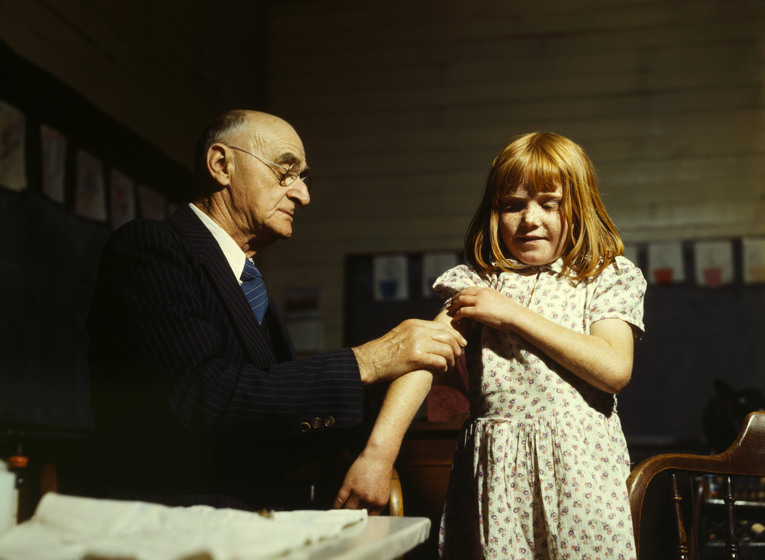
A child receives a typhoid vaccination
https://upload.wikimedia.org/wikipedia/commons/b/be/Typhoid_inoculation2.jpg
Pertussis: When to test and treat
- Anyone who has been exposed should get tested
- Sick babies: sepsis or apnea
- Think about if < 4 mo
- If strongly suggestive history, consider testing
Influenza:
- Earliest 6 mo (IM only)
- 1st year: need 2 doses (1 month apart) for priming, improved antibody response
- Immune response varies yearly, cannot count on complete protection
- Test within 72 hours of illness and going to treat/severe illness
Ohio Statewide Immunization Information System: if you're not sure if they have been vaccinated.
Take Away Points:
- Immunization status can help you risk stratify patients. If unsure, use caution and consider blood cultures when the source of infection is unclear.
- Ask the parent if they are vaccinated but if uncertain contact the pediatrician or use the statewide information system.
- A good question to ask is how old was your child when they last had shots?


