Grand Rounds Recap 4.19.2017
/R4 SIM with Drs. Axelson, Grosso, Richardson and Riddle: Tox Tox Everywhere
Oral Boards
Case #1: A 42 yo male presents a few hours after spilling wheel cleaner on his hands. Diagnosis is hydrofluoric acid burns to the fingers. Treatment with calcium gluconate paste.
Debriefing Points:

- HF is a common ingredient in metal cleaners and glass etching solutions
- Large (%BSA) exposures or limited exposures to high concentrations (> 50%) can cause systemic symptoms due to fluoride binding with Ca, Mg, hyper K, hypo Ca, ventricular dysrhythmias
- Treatment for low concentration/local = Calcium gluconate paste, titrated to pain relief. More severe local injuries may require intra-arterial calcium infusion
- In arrest, may need many doses of CaCl, NaHCO3
Case #2: An 19 yo male with a history of MRSA infections presents c/o what he thinks is a boil on the back of his leg. He had been triaged to the lobby, but during his two hour wait developed severe leg pain, abdominal cramps and vomiting. Diagnosis – black widow spider envenomation. Treatment – antivenin.
Debriefing Points:
- Diagnosis is clinical … labs don’t help
- Almost never fatal, but pain is often resistant to opioids and benzos and can last hours to days
- Antivenin usually relieves symptoms with one 2.5 mL dose (takes 30-60 minutes)
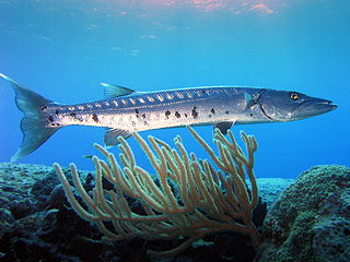
Case #3: A 23 yo female who ate fish on vacation in Florida presents with paresthesias and reversal of hot cold sensation. Diagnosis: Ciguatera poisoning. Treatment: Supportive Care +/- Mannitol.
Debriefing Points:
- Diagnosis is clinical … labs don’t help (but can r/o electrolyte disturbances)
- Source is a heat-stable toxin found in reef fish (from algae they eat)
- Starts with GI distress 1-6 hours after eating the fish, then the neurologic symptoms occur 24-72 hours later
- Mannitol thought to help by decreasing neuronal edema (studies mixed).
- Is a reportable disease in many sea-side communities (due to public health risk)
- Relapse possible over next 3-6 months – guidelines suggest patients should avoid eating any kind of fish, drinking alcohol or becoming dehydrated
Case #4: Chronic Digoxin Toxicity: 73yo female with cc of weak and dizzy. Hyperkalemic, AKI, and elevated Dig level.
Debriefing Points:
- Digoxin toxicity can be acute (taking a large overdose) or chronic (often precipitated in elderly via medication interactions, including diuretics)
- Acute overdoses tend to have higher increase in Digoxin level compared to chronic
- Acute overdoses correlate with degree of hyperkalemia, whereas chronic overdoses can be associated with hypo, normo, or hyperkalemia
- Characteristic EKG findings include ventricular and atrial tachycardias, AV blocks, as well as the classic “scooped ST segments” and flattened T waves
- Treatment for digoxin toxicity is Digifab, monoclonal antibody to digoxin. A single 40mg vial of digifab binds ~0.5mg of digoxin. This can be calculated in acute ingestion, but empiric dose of 5-6 vials is generally given in chronic toxicity, with an empiric dose of 10 vials given in acute toxicity.
- Classically in digoxin toxicity patients describe seeing yellow/green halos around objects: xanthopsia
Simulation
40yo psychiatric patient presents altered, tachypneic, and agitated after an acute ASA ingestion. Eventually the patient seizes and demands intubation, requiring ventilator management in the setting of a severe ASA overdose.
Sources of salicylate exposure:
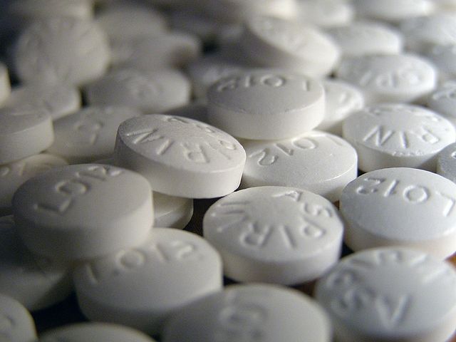
- Salicylates are found in hundreds of over-the-counter medications and in numerous prescription drugs
- Pepto-Bismol, a common antidiarrheal agent, contains 131 mg of salicylate per tablespoon
- Methyl salicylate (e.g., oil of wintergreen) - One teaspoon of 98% methyl salicylate contains 7000 mg of salicylate - more than 4x the potentially toxic dose for a child who weighs 10 kg
Pathophysiology: The classic VBG is a mixed respiratory alkalosis and anion gap metabolic acidosis:
- Anion gap metabolic acidosis results from the uncoupled oxidative phosphorylation intracellularly which limits production of ATP and increases lactate production, leading to ketosis
- Respiratory alkalosis results from a direct stimulation of the respiratory center, leading to hyperventilation
Determining the severity of salicylate ingestion:
- In acute overdose, morbidity rate is 16%, and mortality is 1%
- Prognosis is worse for chronic overdose/exposure because they are often missed
- The following 4 categories are helpful for assessing the potential severity and morbidity of an acute, single event, nonenteric-coated, salicylate ingestion:
- Less than 150 mg/kg - Spectrum ranges from no toxicity to mild toxicity
- 150-300 mg/kg - Mild-to-moderate toxicity
- 301-500 mg/kg - Serious toxicity
- Greater than 500 mg/kg - Potentially lethal toxicity
Organ System Effects:
- Psychiatric:
- The chronic ingestion of salicylates may produce the appearance of anxiety with its associated tachypnea, difficulty concentrating, and hallucinations.
- Pulmonary:
- Salicylates cause both direct and indirect stimulation of respiration. A salicylate level of 35 mg/dL or higher causes increases in both rate (tachypnea) and depth (hyperpnea) of respiration. Salicylate poisoning may rarely cause noncardiogenic pulmonary edema (NCPE) and acute lung injury in pediatric patients.
- Cardiovascular:
- Tachycardia, Hypotension
- Neurologic:
- Salicylates are neurotoxic; this initially manifests as tinnitus. CNS toxicity is related to the amount of drug bound to CNS tissue. It is more common with chronic than acute toxicity. Acidosis worsens CNS toxicity by increasing the amount of salicylate that crosses the blood brain barrier and increases CNS tissue levels.
- The final common pathway is seizure, coma, death
- Gastrointestinal:
- Nausea and vomiting are the most common toxic effects. This can be caused by CNS toxicity or by direct damage to the gastric mucosa.
- Dermatologic:
- Diaphoresis is a common sign in patients with salicylate toxicity.
Diagnostic Testing:
- Chemistry panel
- Repeat as needed
- Serum salicylate level
- Every 2 hours until the salicylate level falls to less than 30 mg/dL
- Serum levels determined less than 6 hours postingestion (acute overdose) do not rule out impending toxicity
- In cases of chronic salicylism, measured toxic levels may be only 30-40 mg/dL
- Acute overdoses are often symptomatic at salicylate concentrations higher than 40-50 mg/dL
- Patients with salicylate concentrations approaching or exceeding 100 mg/dL usually have serious or life-threatening toxicity
- Urinalysis: Monitor and maintain an alkaline urine pH every 2 hours during alkalinization therapy. Maintain a urine pH of 7.5-8.
- Monitor glucose levels closely. Initial hyperglycemia may give way to hypoglycemia.
- Obtain hepatic, hematologic, and coagulation profiles for patients with clinical evidence of moderate-to-severe toxicity
- Chest x-ray is indicated if evidence of severe intoxication, pulmonary edema, or hypoxemia is present
- Consider an abdominal x-ray if an aspirin concretion is suspected
Treatment:
- Decontamination
- Gastric lavage may be beneficial in severe, large volume overdose
- Repeated doses of charcoal may enhance salicylate elimination and may shorten the serum half-life. Most experts strongly recommend this for patients with a serious ingestions
- Administer lactated Ringer or isotonic sodium chloride solution for 1-1.5-cc/kg/h UOP is established.
- Alkalinization of urine creates a gradient that attracts the unionized salicylate from the brain tissue into the blood stream and then into the urine. The goal is to keep salicylates away from brain tissue, move them to the urine, “trap” them in the urine in an ionized state, and then excrete them.
- When the urine pH increases from 5 to 8, salicylate renal clearance increases 10-20 times.
- Consider this treatment if the salicylate level is higher than 35 mg/dL.
- Give a single IV bolus of NaHCO3 at 1-2 mEq/kg.
- Mix 3 ampules of NaHCO3 in 1L of D5W. Add 20-40 mEq KCl.
- Administer a constant infusion at 1.5-2.5 mL/kg/hr to produce a urine flow of 0.5-1 mL/kg/hr. Closely monitor the serum electrolytes (K), cre, HCO3, and urine pH. Maintain the urinary pH between 7.5-8.
- The urinary excretion of salicylic acid is dependent upon adequate serum potassium due to a H+/K+ exchange.
- Dialysis is recommended for patients with severe salicylate poisoning who are altered, have respiratory distress, cannot handle a fluid load, severe academia (<7.2), or not responding to current therapy. The Extracorporeal Treatments in Poisoning (EXTRIP) Workgroup recommended extracorporeal treatment for high salicylate concentrations regardless of signs and symptoms (>100 mg/dL) or lower thresholds for renal impairment (>90 mg/dL]).
Disposition:
- Admit patients with major signs and symptoms to an ICU.
- Patients with accidental ingestions of less than 150 mg/kg and no signs of toxicity can be discharged after 6 hours post ingestion.
Take-Home Points:
- Salicylates uncouple oxidative phosphorylation intracellularly – a key point in understanding the pathophysiology of salicylism.
- Salicylates directly stimulate the medullary respiratory center of the brainstem resulting in tachypnea, classically causing an early pure respiratory alkalosis (centrally mediated), followed by an increased anion gap metabolic acidosis with continued hyperventilation.
- Salicylate-intoxicated patients have rapid respiratory rates and large tidal volumes (increased minute ventilations) that are difficult to maintain with a ventilator. If not maintained, acidemia worsens, and death may ensue.
- Treatment principles include stabilizing the ABCs, limiting absorption, enhancing elimination, correcting electrolyte/metabolic abnormalities, and providing supportive care.
- Alkalinize the urine early to trap the salicylate molecule in the ionized form in the urine and facilitate excretion by giving intravenous sodium bicarbonate boluses and a bicarbonate infusion at twice maintenance rate.
- Replace potassium to maintain the potassium-hydrogen exchange in the renal tubules and to maintain urinary alkalinization.
- Assess serial salicylate levels every 1-2 hours, along with serum glucose, potassium, bicarbonate, and creatinine levels until the salicylate levels are less than 30 mg/dL.
- Hemodialysis is effective for salicylate removal and may be required continuously to remove tissue stores.
R2 Case Follow-up with Dr. Randolph
Young male with a PMHx of DMI who presents to OSH in DKA with chest pain, with concern for pericarditis on EKG in addition to his DKA initially. Another EKG shows an anterior/lateral MI and has 99% LAD, developed florid heart failure (EF 10% with LV dilation), Impella placed and pt transferred for eval for LVAD. LVAD placed and A1C found to be 13.
Pericarditis:

- L sided, positional CP
- EKG Changes: Percarditis (convex J point, distribution of ST elevations, no hyperacute T waves, less QT prolongation)
- Stage 1: ST elevations, PR depression
- Stage 2 (first week): ST elevations normalize
- Stage 3: T wave inversions
- Stage 4: Chronic pericarditis
- High risk for admission:
- Fever and leukocytosis
- Cardiac tamponade
- Large pericardial effuse
- Immunosuppressed state
- Therapy with vit K
- Acute trauma
- Failure to respond to therapy
- Elevated troponin
Type 1 Diabetics
- Complications:
- Hyperglycemia, DKA
- Hypoglycemia
- Growth issues
- Kidney failure
- Cardiac complications:
- Known higher mortality than DMII
- Mortality of 35% at 40 yo compared to 4% in normal population
- Women 8x more likely to have cardiac disease, Men x4
- Decreased SV, Contractility, Stroke volumes
- Increased Total cholesterol, lipids
- Roughly correlated with A1C
- High risk: >12%
- Moderate risk: >9.7%
- Mild risk: >7%
- CONFIRM Study:
- 28% had >50% stenosis asymptomatically on CT scan
- 70% had coronary abnormality
- Known higher mortality than DMII
Pediatric Pain Management with Dr. Spigner
Topical Anesthesia:
- LET (lidocaine, epinephrine, tetracaine)
- Best for face and scalp
- Caution w/ mucous membranes
- Takes 20-30min, lasts 45-60min
- Gel>aqueous
- No evidence for weight based dosing, MedHb rare but caution in <1mo
- EMLA (Lidocaine + Prilocaine)
- Takes 60min, lasts 2-4 hours
- Intact skin
- LMX-4 (Lidocaine)
- Takes 30min, lasts 60min
- Intact skin
- Summary:
- They all work
- LET appears superior for lacerations and fast working
- EMLA/LMX may be better for end-arterial circulation
- Topicals have best efficacy for scalps and face
Intranasal Medication
- Fentanyl: Just as good as morphine when used at 2mcg/kg dose for Abscess I&D, long bone fracture
- 1 mcg/kg/nostril
- Double the dose for nebulized fentanyl
- Just as good IV as nebulized/nasal
- Ketamine: not great data
- No trials for fractures and no direct comparisons
Non-opiate medication
- Will it help if I alternate ibuprofen & acetaminophen?
- In a systematic review: combination was slightly superior to either alone
- Risk: complex dosing regimen, danger of APAP OD
- Why can't I use ibuprofen in children <6mo?
- Not FDA approved
- Concern for renal dysfunction and GI bleeding
- Data is not great, more theoretical than practical
Clinical Capstone with Dr. Derks: Physician Burnout
Burnout: emotional exhaustion and a reduced sense of personal accomplishment associated with prolonged occupational stress
Physical exhaustion, poor judgement, cynicism, guilt, feelings of ineffectiveness, and a sense of depersonalization in relationships with co-workers and patients
Residency itself may induce depression (15-20% of those people get better on their own)
Cost of the Problem: 30-50K per new FTE for the ED, higher in other specialties; worse physician health means more back up activation, more tests and studies ordered per patient, worse patient care. It is in all of our best interest to identify it early, identify it in others and seek treatment early
Taming the SRU with Dr. Miller: Hypothermic Cardiac Arrest
Presumed accidental hypothermic arrest with 2 hours of exposure, initial core temp 69. Intermittent ROSC with ECMO consulted but given persistent acidosis to 6.6 and inability to achieve ROSC, ECMO was not pursued.
1 Study of 10 patients with cardiac arrest from accidental hypothermia showed ECMO to help with survival.
ECC v ECMO: ECC is for 2-3 hours, direct diffusion of gas into the blood. ECMO can prolong use, gas diffusion across a membrane
ECMO advantages:
- Cannulation can be performed percutaneously outside the OR
- Requires lower levels of anticoagulation and can be used without systemic anticoagulaiton
- Portable
- Run for days
- High rates
Asphyxia-related case of hypothermia greatly increased mortality such as primary drowning or suffication (85% v 55%)
Predictors of poor outcome in hypothermic arrest:
- Primary asphyxia
- pH < 7
- K > 10
ECMO is recommended by the European Resuscitation Council for cardiovascular collapse from hypothermia.


