Grands Rounds Recap 4.26.2017
/M&M - MSK US - UGIB Case - CSF Analysis - CPC
Morbidity and Mortality with Dr. McKean
Cases 1 & 2: 2 patients with chest pain who presented with concerning stories, both with previous normal stress testing, normal EKGs, and initial troponin which are all normal. Both were sent to the cath lab with >99% stenosis of coronary arteries.
Learning Points:

- Impact of a negative prior stress test on EM physician decision in ED patients with chest pain
- Adverse outcomes were the same for those with normal stress test v no stress test at all
- Impact of stress test on 30-day cardiovascular for low-risk patients with chest pain
- Outcomes were the same for those admitted, observed and stressed, or sent home
- Predictive value of the exercise EKG for MACE in patients with presented with CP to the ED
- Low-risk CP patients (HEART score <4) had high rate of false positive stresses, very low rate of MACE regardless of whether they were stressed or not.
- Prognostic Implications of angiographically normal and insignificantly narrowed coronary arteries
- The more coronary artery disease on the cath, the worse they did over the next 10 years
- Clinical restenosis after coronary artery stent placement
- This should take at least ~6 months to happen, physiologically (this is different than in-stent thrombosis)
- Within 12 months, 12% in-stent restenosis, 16% had any MACE (death or need for re-catheterization)
- Risk factors for in-stent restenosis:
- Female Gender
- Diabetes
- Post-procedure stent diameter
- Stent length
- Diabetes
- Multi-vessel disease
- Female Gender
Case #3: Patient found down in the bathroom, initial concern for stroke because she only moves her RUE & RLE to pain. She is intubated on scene, mild metabolic acidosis on arrival, leukocytosis, normal parenchyma on head CT but shows left sided otomastoiditis. Upon admission, gets LP that shows frank pus and grows GPC, thought to be pneumococcal. Got a mastoidectomy and antibiotics
- Mastoiditis: intracranial complications in 27.4% in one retrospective review
- 15/17 with meningitis
- Pneumococcal meningitis:
- AMS in 90% of patients
- Focal neurological deficit in 30-40% of patients
- Check out the EBM pathway for recommendations on those who need CT before LP and antibiotics recommendations
Case #4: IVDU with arm pain on initial visit, mild swelling of his arm, normal vitals, normal xray, I&D performed with small amount of blood. Re-presents with HR 200, febrile to 101, RR of 52, not initially hypotensive but with crepitus on exam of RUE and then gas on x-ray. Got a glenohumeral disarticulation for source control.
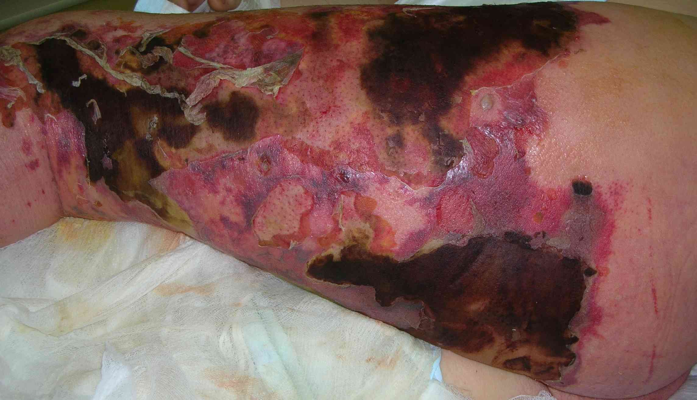
- In one retrospective review of neccotizing fasciitis:
- 80% of patients were IVDU
- Inter-rater reliability for physical exam findings in those with necrotizing fasciitis was very poor
- Only 57% were given the diagnosis of necrotizing soft tissue infection
- Those who were admitted to a surgical service tended to do better than those who were admitted to a medical service
- Pain out of proportion
- When compared to severe cellulitis, "strong" was used to describe pain more often in necrotizing soft tissue infections
- Management:
- Surgery
- Pip/tazo, Vancomycin, and Clindamycin
- Clindamycin is used for "The Eagle" effect, which is a toxin-neutralizing mediated effect
Case #5: Pt fell off a ladder ~20ft, hypothermic with a GCS 14 in the field, RR 38, pulse 100 and normal BP, complaining of right hip pain and back pain. Acidotic, elevated lactate, INR >1.4. Pelvic fractures noted on x-ray. Large retroperitoneal hematoma, cardiac arrest in the scanner, got product, deteriorated to asystole without cardiac activity.
- Keep in mind the triad of death in trauma:
- Acidosis
- Hypothermia
- Coagulopathy
- Study showing with femoral v subclavian line placement during CPR:
- Subclavian lines are safe during CPR but is operator dependent
- In trauma, some data showing femoral lines do not have the same increased infection complications shown in medical patients.
Musculoskeletal US with Dr. Brenkert
Tips:
- Use the opposite side
- Dynamic evaluation
- Lots of gel
- Compression
- Water bath or stand off pad
- And more gel
Anisotropy- angle of insonation: something is falsely dark. Weak echo detected by probe because of the angle of reflection, not because of the make-up of the structure. Fan your probe back and forth to correct for this.
The Hip: sensitivity 85%, specificity 93%

- Technique:
- Patient supine
- Ideal leg positioning: knee slightly flexed, internal rotation
- Linear probe (curved in adults)
- Marker towards the head
- Probe in line with the femoral head/neck
- Lateral to the femoral bundle
- Image both hips
- Anatomy:
- Get a view of the ilioposais, sartoruis, quadraceps above the femoral head and neck
- Abnormal
- Measure the anechoic fluid at the largest thickness across from the femoral neck:
- >5mm in all ages is abnormal
- >2mm difference from contralateral hip is also abnormal
- Measure the anechoic fluid at the largest thickness across from the femoral neck:
- Consider US guidance for hip aspiration
- In plane technique
- Spinal needle
- Same probe orientation
- Consider with the Kocher Criteria (3/4 gives 93% sensitivity for septic arthritis)
- Non-weight bearing
- Temp >101.3
- ESR >40
- WBC >12k
The elbow: 2yo presents holding flexed elbow after an unwitnessed injury
- Supracondylar v Nursemaid's:
- POCUS on 130 kids already getting x-rays for suspicion of fxs
- US had sensitivity 98%, specificity of 70%
- Could reduce x-rays by 48%
- POCUS on 42 pts with RHS clinically
- 80% had a normal fat pad
- Technique: Over the olecranon fossa
- Look at the fad pad: Does it raise up out of the olecranon fossa?
- if yes, then concern for fracture
- Lipohemarthrosis is also concerning for fracture
- Look at the fad pad: Does it raise up out of the olecranon fossa?
- In those with diagnostic uncertainty:
- Posterior fat pad or lipohemarthrosis: get an x-ray
- Normal: try to reduce
Shoulder dislocation:
- 100% sensitive and specific compared to radiology both pre and post reduction.
- With 30 minutes of education and obtaining a single posterior view, 100% sensitive, spec, NPV, & PPV.
- Technique:
- Linear or curved
- Start lateral, go posterior and find the glenohumeral head and its articulation with the fossa (or lack there of)
- Abnormal:
- Humeral head sits closer (posterior dislocation) to the probe or farther away (anterior dislocation) from the probe
Consider 5minsono.com for refreshers on most US techniques.
R4 Case Follow-up with Dr. Mudd: Transfusion for Upper GI Bleeding
Case: Elderly female with OHCA from Upper GI bleed who got 9/9 in the ED and then went to the OR with Gen Surgery and GI for source control within an hour of being in the ED.
Transfusion strategies for Upper GI bleed

- Restrictive transfusion group got transfused for HgB <7, Liberal transfusion group got transfused at HgB <9. All got IVF resus until H/H returned
- Rockall scores of 5 (25% rebleed risk, 11% mortality)
- 1/3 of the patients in shock on arrival
- Restrictive strategy group had a mortality benefit (HR 0.55, NNT 25)
TXA for Upper GI Bleed: NNT of 7 for a mortality in older studies, however this is being re-looked at via a RCT studying being done now
R1 Clinical Diagnostics: CSF Analysis with Dr. Ventura
Check out Dr. Ventura's asynchronous post on CSF analysis here.
Neutrophil to lymphocyte ratio: ANC/ALC in both blood and CSF were useful for differentiating viral from bacterial meningitis
CSF: NLR of 2 increased your risk of bacterial etiology 3x
- With NLR >/= 2 and NC >/= 287 OR 79.5
CSF lactate: produced by bacterial anaerobic metabolism, not affected by your serum lactate. Cutoffs of >27mg/dL.
Criteria for screening for HSV:
- HIV Positive
- Hx of organ transplant
- Age <2yo
- CSF WBC >5
- CSF protein >50
CSF in SAH
- Xanthochormia: presence requires enzymes so differentiates between traumatic tapped
- 100% of confirmed SAH had xanthochromia between 12h-14d
- RBC clearing: not always a reliable marker of the difference between traumatic and SAH, so use with caution
Guillian-Barre
- Brighton Criteria: there is variation of cytoalbuminologic dissociation
- Depends on when in the disease process you perform the CSF
CPC Dr. Whitford vs Dr. Stettler
Case: Teenage patient with type I DM found down, pH 6.9, HCO3 5 AG 19 FSBS 189, somnolent with non-focal neurological exam, normal head CT.
- Primary non-anion gap metabolic acidosis
- Primary respiratory acidosis
- Primary (mild) anion gap metabolic acidosis
Diagnosis: Type 1 Renal Tubular Acidosis secondary to ibuprofen ingestion
Delta Gap: Change in AG/Change in HCO3
- <0.4 is a primary non-anion gap acidosis
- >1.0 primary gap acidosis
Delta criticism: Assumes a normal gap (hypoalbuminemia will throw this off)
Non-gap metabolic acidosis:
- H: Hyperchloremia
- A: Acetazolemide
- R: RTA
- D: Diarrhea
- U: Urinary Fistula
- P: Pancreatic/Enteric fistulas
Can use Urine Anion Gap to diagnose where the loss is coming from:
- + means kidney lossses
- - means gut losses


